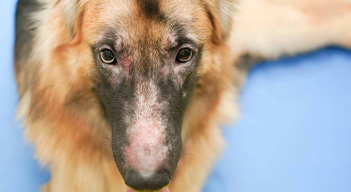
Dogs suffering from atopic dermatitis have an inherited predisposition to develop antibodies to environmental allergens and upon contact with these allergens develop clinical signs. There is a basic defect in the control and regulation of the immune system. Atopy is thought to affect between 3 and 5% of the canine population and has a strong breed predilection. Predisposed breeds include West Highland White Terriers, Cairn Terriers, Scottish Terriers, Wirehaired Fox Terriers, Dalmatians, Pugs, Lhasa Apsos, Irish Setters, Boston Terriers, Golden Retrievers, Boxers, English Setters, Labrador Retrievers and Miniature Schnauzers. The disease classically starts in young dogs (1-2 years of age but as young as 3 months). About 80% of atopic dogs initially show the signs seasonally, but many eventually progress to have continual clinical signs.
Clinical Signs
Itching is typically the hallmark of the disease. The skin lesions are usually associated with self trauma, secondary skin infections and abnormal grease production. Ear infections are seen in 50% of patients and can sometimes be the only sign. Other typically involved sites include the face, ears, feet, lower abdomen and chest, armpits and around the anus. Complicating secondary infections of the skin with bacteria or yeast organisms are extremely common.
Diagnosis
Atopy is diagnosed by history, physical examination and exclusion of other possible causes such as food allergy, flea allergy, contact allergy, sarcoptes mange mites, skin infections etc. A seasonal dermatitis in winter points to allergies to house dust mites (very common). If the dermatitis is seasonal in spring or early summer grass and tree pollens or mould spores may be the problem. Summer and autumn dermatitis may indicate either weed pollen or flea bite allergy. Many patients will show signs for an extended period. In contrast to humans, allergies in dogs tend to become worse with age as they develop allergies to a wide range of allergens.
Intradermal Skin Testing
Intradermal skin testing is considered to be the test of choice to identify the offending allergens and serve as a basis for formulating an allergy vaccine. The patients are sedated using a reversible sedative Domitor in combination with a pain killer butorphanol which is a very safe combination. An area the size of a postcard is clipped on the side of the chest and three rows of felt pen dots are used as reference points. Thirty six antigens are injected in to the skin above and below each dot. Skin test reactions peak between 15-20 minutes and are graded according to their size and redness.
Drugs may influence skin test reactivity. The following withdrawal times prior to testing are necessary to prevent interference with the test results:
- Injectable steroids 6-8 weeks
- Prednisone 4-6 weeks
- Antihistamines 2 weeks
- Topical treatments such as ear & eye drops 2 weeks
Antibiotics and shampoos do not interfere and can be used up to the day of the test.
Treatment
Unfortunately avoidance of the offending allergens is rarely possible. When treating animals with allergies, it is important to understand the hypersensitivity threshold concept. A certain allergic load may be tolerated by an individual without clinical signs, but a small increase may push the animal over the threshold and initiate clinical signs. Controlling only one out of several allergens may be enough to bring the animal in to remission by pushing it back so that it is not allergic enough to itch. For example, good flea control in a dog with house dust mite and flea allergies may stop the itching or ear infections if the dog is below that magic threshold.
Hyposensitisation (“allergy shots”) is currently considered the treatment of choice for most atopic dogs. It has been reported to be effective in 60-80% of atopic dogs. The exact mechanism of these injections is still unknown. An allergy vaccine is made up using the antigens identified by intradermal testing. The owner is taught how to administer the injections. They are initially given every 3 days while the dose of antigens steadily increases. After 6 weeks the interval between injections is increased. Maintenance injections are generally given every 2-4 weeks depending on the response. They continue for at least 2 years and often for the rest of the dog’s life.
Numerous studies in animals have shown some value in supplementation with essential fatty acids (EFAs) in allergic skin disease. While an excellent response (no other medication) is achieved in only 10-20% of cases, as an adjunct therapy (together with hyposensitisation, or antihistamines and shampoo therapy) the use of drugs like prednisone can either be minimised or even avoided in many cases. The best results are achieved by feeding foods with the specific ratio and amount of the omega 3 and omega 6 EFAs scientifically added to the diet. We recommend either Eukanuba Dermatosis or Hill’s J/D. Where there are financial constraints, sunflower or safflower oil can be added to the diet at a rate of 1 tablespoon per 10kg body weight daily (start gradually to avoid diarrhoea)
